Venography: Types, Procedure Steps and Recommendations
Venography is a method that uses X-rays to examine blood vessels and diagnose disorders such as varicose veins and blood clots.


Venography is a method that uses X-rays to examine blood vessels and diagnose disorders such as varicose veins and blood clots.
- What is Venography and Why is it Important for Varicose Veins?
- Types of Venography: Which Method is More Suitable?
- Venography has various types, performed according to the specific problem and the physician's diagnosis:
- Applications of Venography in Venous and Vascular Diseases
- Venography is also used in diagnosing venous and vascular diseases and disorders including:
- What Should Be Done Before Venography?
- Steps of Performing Venography
- Before the venography the patient may be asked to wear a special gown and remove metal objects such as jewelry or glasses. Then they lie down on a special table or bed.
- Advantages of Venography for Varicose Veins
- According to research the advantages of this method include:
- Post-Venography Care and Measures
Venography is a specialized imaging method using X-rays, employed to diagnose venous problems such as varicose veins, deep vein thrombosis (DVT) and venous insufficiency.
What is Venography and Why is it Important for Varicose Veins?
Venography (or Phlebography) is a medical imaging technique that uses a special contrast agent and X-rays to provide a detailed image of the body's venous system. This method is recognized as the "gold standard" for diagnosing many venous diseases especially when other non-invasive methods do not provide definitive results. According to authoritative sources such as the Radiological Society of North America (RSNA) and the American College of Radiology (ACR), venography plays a vital role in accurately diagnosing conditions like deep vein thrombosis (DVT), venous insufficiency, congenital vascular anomalies and evaluating veins prior to surgical procedures.
Types of Venography: Which Method is More Suitable?
Venography has various types, performed according to the specific problem and the physician's diagnosis:
Standard (Traditional) Venography: This classic method involves injecting a contrast agent into a vein and recording X-ray images. According to guidelines from the American Heart Association (AHA), this method is widely used for assessing venous blood flow and identifying blood clots.
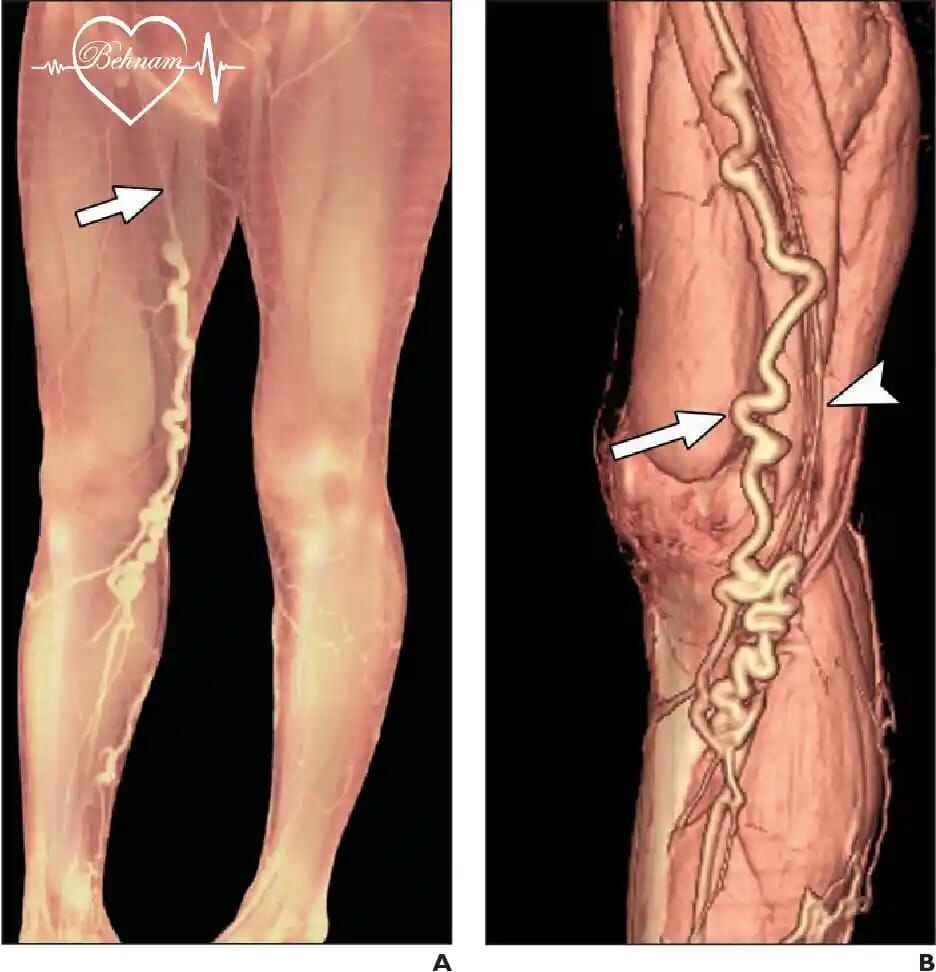
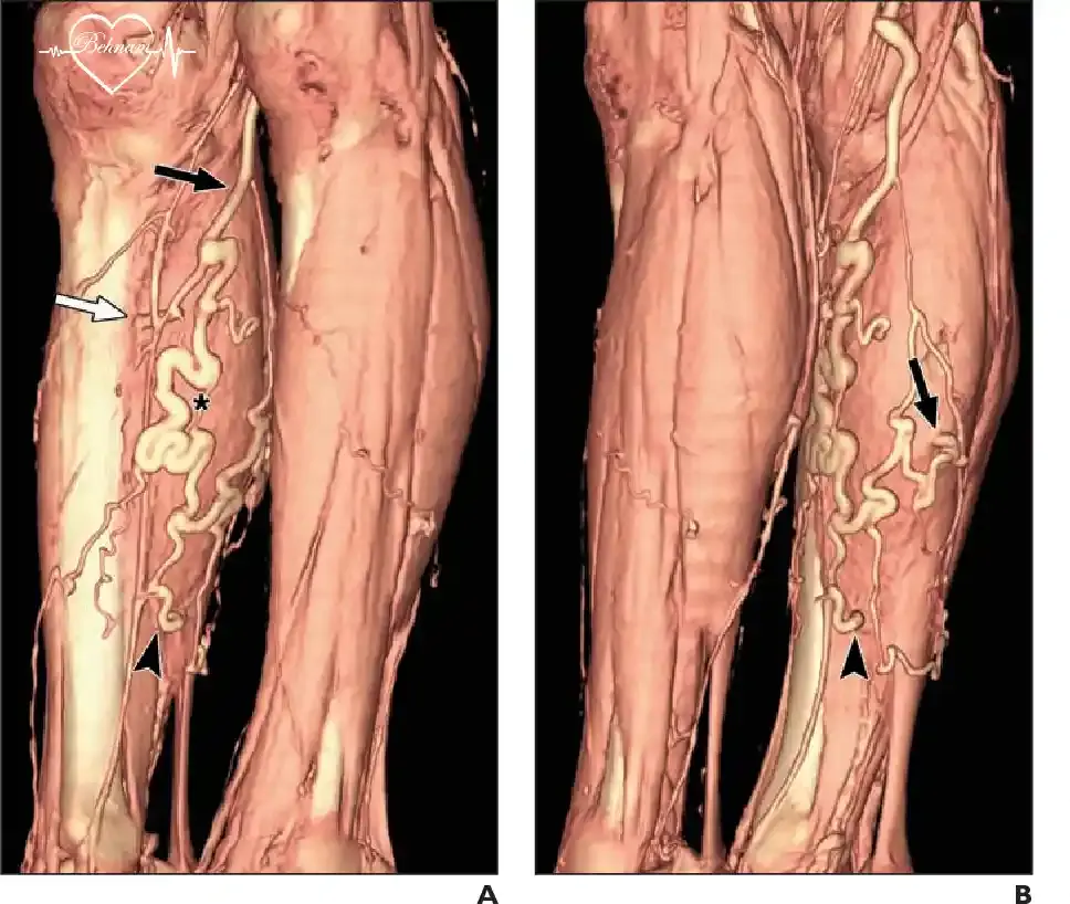
CT Venography (CTV): In this advanced technique after injecting the contrast agent a CT scanner is used to produce detailed three-dimensional images of the venous system. Studies indicate that CT venography has high sensitivity in diagnosing DVT or deep vein thrombosis.
MR Venography (MRV): MR venography is performed without using X-rays, utilizing a strong magnetic field. This method is a safe option for patients sensitive to radiation or iodinated contrast agents because it does not require radioactive injection and can provide precise images of the body's veins and blood vessels.
Applications of Venography in Venous and Vascular Diseases
Venography is also used in diagnosing venous and vascular diseases and disorders including:
Diagnosing Deep Vein Thrombosis (DVT): DVT is one of the most dangerous venous diseases that can lead to pulmonary embolism and death. Venography can accurately identify the presence, size and exact location of the clot.
Evaluating Varicose Veins and Venous Insufficiency Using Venography: Venography provides a complete map of the leg venous network and helps the physician:
Identify the source of reflux (backflow of blood).
Assess the function of venous valves.
Choose the best treatment method for varicose veins such as laser, radiofrequency, sclerotherapy or surgery.
Before performing complex operations like venous bypass surgery, venography provides vital information about the anatomy and functional status of the veins.
What Should Be Done Before Venography?
Fasting for 4-6 hours before the procedure (as per doctor's orders).
Informing about allergies and sensitivities (especially to iodine or contrast agents).
Reporting a history of kidney, heart diseases or diabetes.
Informing about pregnancy or the possibility of pregnancy.
Steps of Performing Venography
Before the venography the patient may be asked to wear a special gown and remove metal objects such as jewelry or glasses. Then they lie down on a special table or bed.
Contrast Agent Injection: Usually done through a vein in the foot or hand. A slight feeling of warmth or numbness may occur which is temporary and will subside.
Imaging: The patient is secured with a safety belt, and images are taken using X-rays from the desired areas. The patient must remain calm and still during this stage.
Image Interpretation: The radiologist analyzes the images. The total duration is usually between 30 to 60 minutes and the patient is typically discharged the same day.
Advantages of Venography for Varicose Veins
According to research the advantages of this method include:
Very high diagnostic accuracy.
Providing precise anatomical information.
The possibility of diagnosing multiple venous problems simultaneously.
Relative availability in medical centers.
Disadvantages and Potential Risks of Venography
Despite its high diagnostic accuracy, venography is considered an invasive method and comes with complications and limitations. Understanding these is essential for informed decision-making by both patient and physician.
Complications Related to Contrast Agent:
A) Allergic and Hypersensitivity Reactions:
Mild to moderate reactions: Hives, itching, nausea, transient feeling of warmth.
Severe reactions: Deep skin swelling, sudden spasm of respiratory muscles and anaphylactic shock which can be life-threatening.
B) Contrast-Induced Nephropathy (CIN) or Kidney Damage:
Definition: Decrease in kidney function within 48-72 hours after injection.
High-risk groups: Patients with pre-existing kidney failure, diabetes, congestive heart failure, dehydration and the elderly.
Prevention: Adequate hydration or drinking sufficient water before and after the procedure, using the minimum volume of contrast agent, alternative options for high-risk patients.
C) Sensitivity to Iodine: The contrast agent used in standard and CT venography usually contains iodine which may cause reactions in individuals sensitive or allergic to this substance.
Complications Related to Intravenous Injection and Vascular Access:
Extravasation or leakage of the agent into surrounding tissue: Can cause pain, swelling, redness, and in severe cases, necrosis or death of skin tissue especially if the solution is highly concentrated.
Thrombophlebitis (vein inflammation): Formation of a clot in the injected vein.
Hematoma or bruising and bleeding at the injection site.
Infection at the catheter entry site (rare).
Adjacent nerve damage (very rare).
Exposure to Ionizing Radiation (in Standard and CT Venography):
Although the radiation dose in a simple venography is low, it is considered a relative contraindication for pregnant women or those suspected of pregnancy (except in vital emergency conditions).
Slightly increased long-term risk of developing cancer especially with repeated procedures.
Disadvantages and Limitations:
Invasiveness: Requires needles, catheters and intravenous injection.
Time-consuming and costly: Compared to Doppler ultrasound, its execution is longer and more expensive.
Limited access to all veins: Evaluating some small or deeply located veins with complete obstruction may be difficult.
Need for patient cooperation: The patient must remain still during imaging.
Limitations in specific patients: Obese patients may not provide high-quality images.
Very Rare but Serious Risks:
Pulmonary embolism during or immediately after the procedure (if an existing clot dislodges).
Reduced blood flow and oxygen to the limb (if injection is accidentally performed into an artery).
Delayed reactions to the contrast agent (late appearance of hives or skin inflammation up to several days later).
Strategies to Reduce Risk and Manage Venography Complications
The following measures can be considered to reduce potential complications and risks of venography:
Accurate pre-procedure evaluation:
Reviewing allergy history (especially to iodine, seafood, previous contrast agents).
Testing kidney function (blood creatinine) in high-risk patients.
Checking for pregnancy in women of childbearing age.
Using modern protocols:
Using new iso-osmolar or low-osmolar contrast agents that have fewer renal and allergic side effects.
Pre-treatment with corticosteroids and antihistamines for patients with a history of allergic reaction.
Substitution with less invasive methods (when possible and as determined by the physician):
Color Doppler ultrasound: First line of diagnosis for DVT and superficial venous insufficiency.
MR venography (MRV): Radiation-free and uses gadolinium-based contrast agents which have a much lower risk of nephrotoxicity or kidney damage.
Adequate hydration: Drinking plenty of water before and after venography to expedite the excretion of the contrast agent.
Comparison of Risks and Benefits of Venography or Phlebography
Venography is recommended when its diagnostic benefit clearly outweighs its risks such as:
High suspicion of DVT with negative or inconclusive ultrasound.
Planning for complex venous surgery.
Evaluating congenital vascular anomalies.
The final decision to perform this procedure should be made after detailed consultation among the treating physician (phlebologist or vascular surgeon), radiologist and the patient themselves, taking into full account the individual's specific conditions.
Alternatives to Venography: Doppler Ultrasound
Color Doppler ultrasound is currently considered the first line of diagnosis for venous diseases and is introduced as a non-invasive, radiation-free and highly accurate method. However venography still holds preference in the following cases:
Inconclusive ultrasound results.
Complex venous anatomy.
Pre-operative evaluation for complex surgeries.
Obese patients where ultrasound has low diagnostic quality.
Venography Results: Interpreting Findings
Normal venography results indicate:
Free flow of contrast agent in all veins.
Absence of filling defects (indicative of clots).
Normal function of venous valves.
No stenosis or obstruction in the venous pathway.
Abnormal findings may include:
Filling defect: Classic sign of a blood clot.
Flow cutoff: Sign of complete venous obstruction.
Reflux (backflow of blood): Sign of valvular insufficiency.
Development of collateral network: Sign of chronic obstruction.
Post-Venography Care and Measures
Drinking plenty of fluids to excrete the contrast agent.
Caring for the injection site and dressing.
Immediately reporting any unusual complication to the physician.
Regular follow-up with a vein specialist (phlebologist) based on the results.
Summary: The Role of Venography in Diagnosing Vascular and Venous Diseases
Despite the emergence of new imaging methods, venography remains a powerful diagnostic tool and the gold standard in many complex venous conditions. The decision to perform this method should be made by the physician, phlebologist, radiologist and vascular surgeon, considering the specific conditions of each patient. Newer techniques like CT venography and MR venography with reduced complications and increased accuracy, promise a bright future for venous imaging. However choosing the appropriate diagnostic method should be based on current scientific evidence and the characteristics of each patient.
Frequently Asked Questions About Venography
Is venography painful?
A brief burning sensation during contrast injection is normal. The entire process is performed with local anesthesia and has tolerable pain.
Who should not undergo venography?
Pregnant women (except in emergency conditions), individuals with severe kidney failure, patients with severe sensitivity to contrast agents, individuals with advanced congestive heart failure.
How accurate is venography?
With an accuracy of over 95%, venography has the highest diagnostic accuracy among venous imaging methods.

Dr. Behnam Vaghefi holds a fellowship in cardiology and vascular diseases, is a specialist in varicose vein treatment, and is a member of the American and European Heart Associations. This article has been written with the review and approval of Dr. Vaghefi, who is the author of several articles and books on cardiology and vascular diseases, and who has years of experience treating patients in his specialized clinic.
Medical Consultation