ECG or electrocardiogram
An ECG (Electrocardiogram) is usually the first test at the beginning of the examination and diagnosis process. It is a quick, easy, non-invasive, painless and radiation-free test.


An ECG (Electrocardiogram) is usually the first test at the beginning of the examination and diagnosis process. It is a quick, easy, non-invasive, painless and radiation-free test.
- When did ECG come into use?
- What is an ECG?
- The ECG Procedure
- What is the most important information an ECG provides?
- Types of ECG Leads
- ECG in ladies
- Normal and normal ECG
- The benefits of electrocardiogram
- What is the Difference Between an ECG and an Echocardiogram?
- Although both methods are used to evaluate the heart, they differ in terms of procedure, evaluation, advantages and limitations.
Electrocardiography (ECG/EKG) is usually the first test in the diagnostic and examination process. An ECG is a quick, easy, non-invasive, painless and radiation-free test performed in a clinic. This non-invasive method is used to record the heart's electrical activity.
When did ECG come into use?
In 1902, the electrocardiograph was invented by the Dutch biologist, Willem Einthoven. Since then with advancements in technology and more sophisticated equipment, this method has been used to diagnose many heart conditions.
What is an ECG?
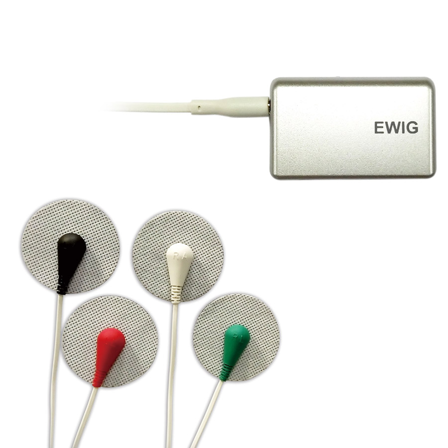
In this method, electrodes are placed on the skin on the chest, arms and legs. The heart's electrical activity is recorded by a device. Essentially the electrical waves generated by the heart are received and displayed as curves on the device. By analyzing these curves the physician obtains information about the heart's size, muscle and rhythm.
The ECG Procedure
An ECG can be performed in a clinic or hospital, following these steps:
Patient Preparation: The patient lies down and clothing from the upper body and chest is removed.
Attaching Electrodes: Electrodes are attached to the patient. The skin at the electrode sites must be dry and clean. These electrodes are placed on the chest, arms and legs.
Recording the ECG: After placing the electrodes on specific areas of the chest, wrists and ankles, the device records the results and generates the corresponding curves. The physician then reviews the results to make a diagnosis. The procedure which usually takes only a few minutes, concludes by removing the electrodes from the patient.
What is the most important information an ECG provides?
Investigating the causes of various types of palpitations and cardiac arrhythmias.
Evaluating heart attack (myocardial infarction) and severe reduction of blood flow to the heart in patients presenting with chest pain.
Diagnosing conditions such as increased thickness and size of the heart wall in patients with hypertension, pulmonary embolism, etc.
Assessing heart failure.
Types of ECG Leads
A standard 12-lead ECG is used, consisting of 6 limb leads (which can be attached to the arms and legs) and 6 chest (precordial) leads. These leads reveal irregularities or arrhythmias in the heartbeat. Sometimes, the information provided by the ECG leads may appear normal. If the physician suspects heart disease, other diagnostic methods may be employed.
Posterior ECG
In a posterior ECG the patient lies on their back (prone). The placement of the chest leads is slightly modified in this method.
ECG in ladies
Care strip from women has no particular complexity and complexity and unlike men who may need to be applied or used to install electrodes because of their hair on their body, electrodes can be easily installed.
patient should be naked from the top of the trunk and remove any ornaments and metals before taking the ECG.
ECG is dangerous in pregnancy?
getting a cardiac and pregnancy during pregnancy and pregnancy has no danger to the mother and fetus because it is a safe and safe procedure. Heart palpitations which are very common during pregnancy as well as mothers with heart disease may need to get a heart band.
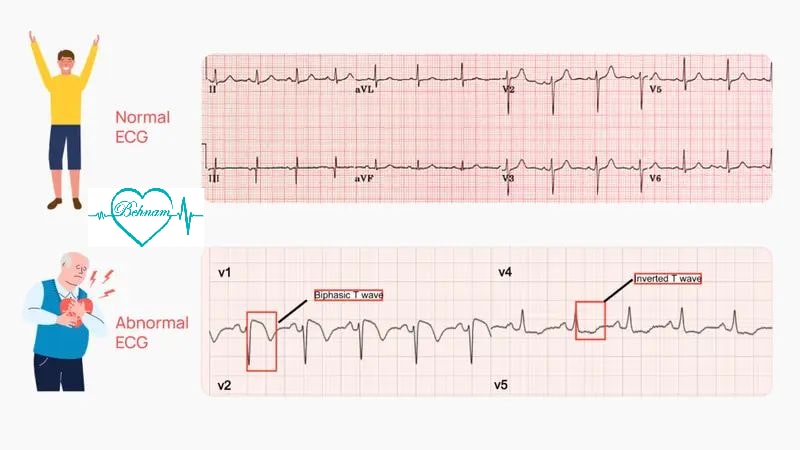
Normal and normal ECG
Natural heart rate is 2 to 5 times a minute. If the patient's heart rate is higher or less than that size, it will be recorded and recognizable. In addition, the heartbeat rhythm should be regular and uniform. The waves and curves displayed by the device must also have a natural shape that will be recognizable by the physician.
ECG disadvantages
This method can have some limitations. For example in some cases, it may not provide the right results due to anxiety in the patient or some heart problems may not be detectable by this method.
The benefits of electrocardiogram
without any pain and bleeding
Easy and Easy to do
do not require any special preparation before doing so like fasting and ...
quick and easy detection
without symptoms
Holter Monitor
A Holter monitor is a small device attached to the patient that records the heart's rhythm for several days. Holter monitors are available in various models, both wired and wireless and can record heart rhythms from 1 day up to 2-3 years. A computer then analyzes the device's data, identifying various cardiac arrhythmias, palpitations and bradycardias (slow heart rates).
When is a Holter Monitor Used?
For individuals who experience occasional palpitations.
For individuals who feel an irregularity in their heartbeat.
What is the Difference Between an ECG and an Echocardiogram?
Although both methods are used to evaluate the heart, they differ in terms of procedure, evaluation, advantages and limitations.
Procedure: An ECG measures the heart's electrical activity using electrodes attached to the body. In contrast an echocardiogram uses high-frequency sound waves (ultrasound) directed toward the heart and their echoes are displayed as images.
Evaluation: An ECG is used to assess heart rhythm, heart damage and the size and thickness of the heart muscle. An echocardiogram evaluates the function of the heart valves, the size of the chambers, detects the presence of blood clots, examines the heart's structure, wall thickness and blood flow.
Advantages: An ECG is a simple, quick method that effectively shows changes in heart rhythm and is less expensive than an echocardiogram. An echocardiogram provides clear and detailed images of the heart, effectively revealing structural and functional problems.
Limitations and Disadvantages: An ECG only shows the electrical activity of the heart and cannot visualize the heart's structure, blood flow or valves. Echocardiography requires a specialist or physician and sometimes image quality may be suboptimal due to factors like patient obesity or thick chest bones.

Dr. Behnam Vaghefi holds a fellowship in cardiology and vascular diseases, is a specialist in varicose vein treatment, and is a member of the American and European Heart Associations. This article has been written with the review and approval of Dr. Vaghefi, who is the author of several articles and books on cardiology and vascular diseases, and who has years of experience treating patients in his specialized clinic.
Medical Consultation