What are the types of ultrasound and tests for diagnosing leg varicose veins?
In addition to the physical examination by a specialist there are now precise and new methods for diagnosing varicose veins. Early diagnosis of varicose veins is considered the first and most important step in treatment.


In addition to the physical examination by a specialist there are now precise and new methods for diagnosing varicose veins. Early diagnosis of varicose veins is considered the first and most important step in treatment.
Varicose veins is a disease that occurs due to disorders in veins and their blood circulation. This condition is most visible in the legs but other parts of the body may also be affected. The presence of swollen and twisted veins is not only unpleasant for patients in terms of appearance but the pain and discomfort this disease causes are also among the problems of varicose veins.
Diagnosis of different types of varicose veins today thanks to advances in technology and medicine includes numerous methods which we will fully examine in this article.
Importance of Varicose Veins Diagnosis
The first and most effective step in treating any disease is its accurate and correct diagnosis because misdiagnosis will lead to irrelevant and incorrect treatment as well. Therefore correct diagnosis not only helps in choosing the varicose veins treatment method but also prevents serious complications of this disease such as blood clot formation, ulcers or infection.
In addition timely and correct diagnosis provides the possibility of simpler and more effective treatments and prevents wasting the time and money spent by the patient.
Methods of Diagnosing Leg Varicose Veins
The most important methods of diagnosing leg varicose veins are:
1- Physical examination
Direct observation by the varicose veins specialist examines the symptoms and signs of varicose veins including the location and size of varicose veins through examination.
Palpation of veins
The doctor evaluates the amount of swelling and hardness of veins by touching them.
Maneuver tests
In these types of tests the position of varicose veins is examined by the doctor while standing or during pressure. For example to evaluate varicocele which is a type of testicular varicose veins the Valsalva maneuver test is used in which the patient is asked to hold their breath and apply pressure. In this case swollen and dilated veins become visible.
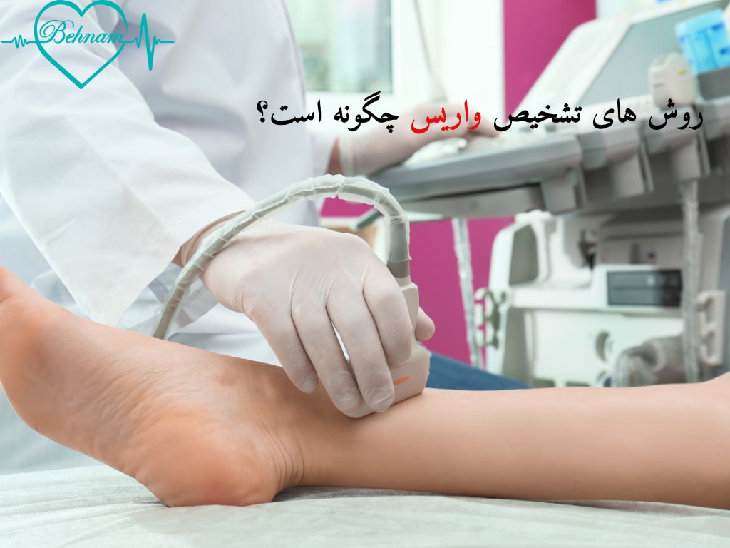
2- Ultrasound and varicose veins diagnostic tests
Doppler ultrasound
One of the most common and accurate methods of diagnosing varicose veins is Doppler ultrasound. In this ultrasound blood circulation in veins is examined using sound waves. This method shows the exact location of varicose veins valve insufficiency in veins and the presence of blood clots.
Doppler ultrasound does not require stitches, incisions or injections and is performed painlessly and without discomfort like regular ultrasound. This type of diagnosis has very high accuracy in providing clear images of veins and blood flow. This method is also used as guidance for treatments such as sclerotherapy or endovenous laser.
It is not time-consuming and does not require special equipment in addition it has lower cost compared to other methods. The patient does not need special preparation and wearing loose and comfortable clothes is sufficient. Given the advantages we mentioned it has the most application in diagnosing varicose veins.
Duplex ultrasound
This method is a combination of Doppler and ultrasound. Like the previous method due to the use of sound waves and being non-invasive it does not create complications or danger and is accessible and low-cost.
Duplex ultrasound is usually performed standing and similar to regular ultrasound and blood flow in veins, the function of vein valves, the location and size of varicose veins or the presence of blood clots and in general the initial diagnosis of varicose veins can be examined through it.
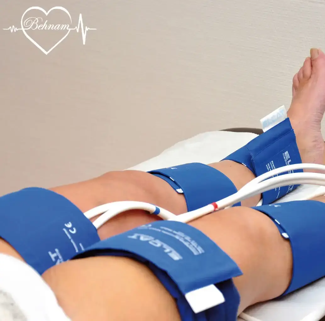
Plethysmography
This method is mostly used to evaluate and diagnose venous insufficiency. Plethysmography examines changes in the volume of a limb. In the case of varicose veins it is used to measure volume changes and the function of the calf muscle pump which plays a role in returning blood to the heart through which information about venous emptying speed, vein blockages and valve function is obtained.
This diagnosis is performed lying down or standing while pressure cuffs (like blood pressure cuffs) are on the leg and sensors are attached to the skin. This method like Doppler and Duplex is non-invasive, painless and without complications and does not require special preparation. The patient may only feel mild pressure from the cuff around the leg.
Plethysmography requires more time and cost and therefore is mostly used alongside other diagnostic methods.
Venography
In this method a contrast agent is injected into the vein and then imaging of veins is done with X-rays. This diagnosis also provides an accurate image of veins and blood flow path and is used for examining complex varicose veins and especially before surgery or evaluating results after it.
In venography the patient lies on their back. After injecting the contrast agent imaging is done with X-rays (live video imaging). Since this method is invasive uses contrast agent and X-rays and has higher cost it is used less than previous methods.
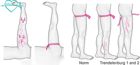
Trendelenburg test
The Trendelenburg test is used to evaluate the function of vein valves. First the patient lies down and then the leg affected by varicose veins is raised. Then the doctor ties a special band or strap called a tourniquet at the top of the thigh. The patient slowly stands again and the doctor examines the speed of blood filling in superficial veins. Rapid or abnormal filling of veins can be a sign of valve disorder and insufficiency.
This test is mostly used for preliminary evaluations and for more accurate diagnosis methods such as Doppler ultrasound should be used.
MRI and CT scan
MRI and CT scan provide accurate images of vein structure and are used in special cases and when there is a need for wider examination of the blood flow network in veins for example in the pelvic area as well.
Summary
Timely and accurate diagnosis of varicose veins plays an effective role in the speed of treatment and preventing its complications. The doctor chooses the appropriate diagnostic method according to the type of varicose veins and the patient's condition. Among the methods we mentioned in this article Doppler ultrasound due to high accuracy and speed, low cost and being suitable for initial diagnosis and evaluations of varicose veins has the most application compared to other methods.
FAQ
1- How is Doppler ultrasound performed and does it hurt?
A little gel is applied on the skin and then the ultrasound probe is moved over the legs. This method is completely safe and painless.
2- Is a blood test necessary to diagnose varicose veins?
In some cases where the doctor suspects a blood clot or another disease a blood test may be needed.
3- How can I diagnose leg varicose veins myself at home?
In case of symptoms such as swollen, raised or purple and blue veins, feeling pain and discomfort in the legs, restless legs syndrome, ulcers and bleeding and skin discoloration you can diagnose leg varicose veins. However due to deep varicose veins you may not have symptoms and to diagnose you must definitely see a doctor.
4- After diagnosing varicose veins should I repeat ultrasound every year?
The answer to this question depends on the severity of the disease and symptoms. If you have new symptoms such as severe swelling or bleeding the diagnosis should be repeated otherwise every 2 to 3 years is sufficient.
Dr. Behnam Vaghefi holds a fellowship in cardiology and vascular diseases, is a specialist in varicose vein treatment, and is a member of the American and European Heart Associations. This article has been written with the review and approval of Dr. Vaghefi, who is the author of several articles and books on cardiology and vascular diseases, and who has years of experience treating patients in his specialized clinic.
Medical Consultation